Circulatory System

http://www.fairview.org/healthlibrary/content/circ.gif

The circulatory system is an organ system that passes nutrients (such as amino acids and electrolytes), gases, hormones, blood cells, , etc. to and from cells in the body to help fight diseases and help stabilize body temperature and pH to maintain homeostasis. This system may be seen strictly as a blood distribution network, but some consider the circulatory system as composed of the cardiovascular system, which distributes blood, and the lymphatic system,[2] which distributes lymph. While humans, as well as other vertebrates, have a closed cardiovascular system (meaning that the blood never leaves the network of arteries, veins and capillaries), some invertebrate groups have an open cardiovascular system. The most primitive animal phyla lack circulatory system. The lymphatic system, on the other hand, is an open system.
http://img2.allposters.com/images/JAG/03-PS121-2.jpg

The main components of the human circulatory system are the heart, the blood, and the blood vessels. The circulatory system includes: the pulmonary circulation, a "loop" through the lungs where blood is oxygenated; and the systemic circulation, a "loop" through the rest of the body to provide oxygenated blood. An average adult contains five to six quarts (roughly 4.7 to 5.7 liters) of blood, which consists of plasma, red blood cells, white blood cells, and platelets. Also, the digestive system works with the circulatory system to provide the nutrients the system needs to keep the heart pumping.

Two types of fluids move through the circulatory system: blood and lymph. The blood, heart, and blood vessels form the cardiovascular system. The lymph, lymph nodes, and lymph vessels form the lymphatic system. The cardiovascular system and the lymphatic system collectively make up the circulatory system.


Surface anatomy of the human heart. The heart is demarcated by:
-A point 9 cm to the left of the midsternal line (apex of the heart)
-The seventh right sternocostal articulation
-The upper border of the third right costal cartilage 1 cm from the right sternal line
-The lower border of the second left costal cartilage 2.5 cm from the left lateral sternal line.

http://www.niaaa.nih.gov/NR/rdonlyres/FD5DD30B-F412-46BF-B407-5421CBD0C8DB/0/271f2.gif
The heart pumps oxygenated blood to the body and deoxygenated blood to the lungs. In the human heart there is one atrium and one ventricle for each circulation, and with both a systemic and a pulmonary circulation there are four chambers in total: left atrium, left ventricle, right atrium and right ventricle. The right atrium is the upper chamber of the right side of the heart. The blood that is returned to the right atrium is deoxygenated (poor in oxygen) and passed into the right ventricle to be pumped through the pulmonary artery to the lungs for re-oxygenation and removal of carbon dioxide. The left atrium receives newly oxygenated blood from the lungs as well as the pulmonary vein which is passed into the strong left ventricle to be pumped through the aorta to the tissues of the body.

History of discoveries A human heart with a visible gun shot wound

A human heart with a visible gun shot wound

The valves of the heart were discovered by a physician of the Hippocratean school around the 4th century BC. However, their function was not properly understood then. Because blood pools in the veins after death, arteries look empty. Ancient anatomists assumed they were filled with air and that they were for transport of air.

Philosophers distinguished veins from arteries but thought that the pulse was a property of arteries themselves. Erasistratos observed that arteries that were cut during life bleed. He ascribed the fact to the phenomenon that air escaping from an artery is replaced with blood that entered by very small vessels between veins and arteries. Thus he apparently postulated capillaries but with reversed flow of blood.

The 2nd century AD, Greek physician Galenos (Galen) knew that blood vessels carried blood and identified venous (dark red) and arterial (brighter and thinner) blood, each with distinct and separate functions. Growth and energy were derived from venous blood created in the liver from chyle, while arterial blood gave vitality by containing pneuma (air) and originated in the heart. Blood flowed from both creating organs to all parts of the body where it was consumed and there was no return of blood to the heart or liver. The heart did not pump blood around, the heart's motion sucked blood in during diastole and the blood moved by the pulsation of the arteries themselves.

Galen believed that the arterial blood was created by venous blood passing from the left ventricle to the right by passing through 'pores' in the inter ventricular septum, air passed from the lungs via the pulmonary artery to the left side of the heart. As the arterial blood was created 'sooty' vapors were created and passed to the lungs also via the pulmonary artery to be exhaled.

The first major scientific understanding of the heart was put forth by the medieval Arab polymath Ibn Al-Nafis, regarded as the father of circulatory physiology.[11] He was the first physician to correctly describe pulmonary circulation,[12] the capillary[13] and coronary circulations.[14] Prior to this, Galen's theory was widely accepted, and improved upon by Avicenna. Al-Nafis rejected the Galen-Avicenna theory and corrected many wrong ideas that were put forth by it, and also adding his new found observations of pulse and circulation to the new theory. His major observations include (as surmised by Dr. Paul Ghalioungui):[13]

1. "Denying the existence of any pores through the interventricular septum."

2. "The flow of blood from the right ventricle to the lungs where its lighter parts filter into the pulmonary vein to mix with air."

3. "The notion that blood, or spirit from the mixture of blood and air, passes from the lung to the left ventricle, and not in the opposite direction."

4. "The assertion that there are only two ventricles, not three as stated by Avicenna."

5. "The statement that the ventricle takes its nourishment from blood flowing in the vessels that run in its substance (i.e. the coronary vessels) and not, as Avicenna maintained, from blood deposited in the right ventricle."

6. "A premonition of the capillary circulation in his assertion that the pulmonary vein receives what comes out of the pulmonary artery, this being the reason for the existence of perceptible passages between the two."

Human Brain, Neuroscience Portal

Illustration of the human brain and skull

The human brain is the center of the human nervous system and is a highly complex organ. Enclosed in the cranium, it has the same general structure as the brains of other mammals, but is over three times as large as the brain of a typical mammal with an equivalent body size.[1] Most of the expansion comes from the cerebral cortex, a convoluted layer of neural tissue that covers the surface of the forebrain. Especially expanded are the frontal lobes, which are involved in executive functions such as self-control, planning, reasoning, and abstract thought. The portion of the brain devoted to vision is also greatly enlarged in human beings.

Brain evolution, from the earliest shrewlike mammals through primates to hominids, is marked by a steady increase in encephalization, or the ratio of brain to body size. The human brain has been estimated to contain 50–100 billion (1011) neurons[citation needed], of which about 10 billion (1010) are cortical pyramidal cells.[citation needed] These cells pass signals to each other via approximately 100 trillion (1014)[citation needed] synaptic connections.

In spite of the fact that it is protected by the thick bones of the skull, suspended in cerebrospinal fluid, and isolated from the bloodstream by the blood-brain barrier, the delicate nature of the human brain makes it susceptible to many types of damage and disease. The most common forms of physical damage are closed head injuries such as a blow to the head, a stroke, or poisoning by a wide variety of chemicals that can act as neurotoxins. Infection of the brain is rare because of the barriers that protect it, but is very serious when it occurs. More common are genetically based diseases[citation needed], such as Parkinson's disease, multiple sclerosis, and many others. A number of psychiatric conditions, such as schizophrenia and depression, are widely thought to be caused at least partially by brain dysfunctions, although the nature of such brain anomalies is not well understood.

Bisection of the head of an adult man, showing the cerebral cortex and underlying white matter
Drawing of the human brain, showing several important structures
Situated at the top and covered with a convoluted cortex, the cerebral hemispheres form the largest part of the human brain .[5] Underneath the cerebrum lies the brainstem, resembling a stalk on which the cerebrum is attached. At the rear of the brain, beneath the cerebrum and behind the brainstem, is the cerebellum, a structure with a horizontally furrowed surface that makes it look different from any other brain area. The same structures are present in other mammals, although the cerebellum is not so large relative to the rest of brain. As a rule, the smaller the cerebrum, the less convoluted the cortex. The cortex of a rat or mouse is almost completely smooth. The cortex of a dolphin or whale, on the other hand, is more convoluted than the cortex of a human.
The four lobes of the cerebral cortex
The bones of the human skull
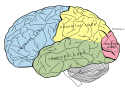
The cerebral cortex is nearly symmetric in outward form, with left and right hemispheres. Anatomists conventionally divide each hemisphere into four "lobes", the frontal lobe, parietal lobe, temporal lobe, and occipital lobe. It is important to realize that this categorization does not actually arise from the structure of the cortex itself: the lobes are named after the bones of the skull that overlie them. There is one exception: the border between the frontal and parietal lobes is shifted backward to the central sulcus, a deep fold that marks the line where the primary somatosensory cortex and primary motor cortex come together.

Topography, Lateralization

Topography of the primary motor cortex, showing which body part is controlled by each zone
Routing of neural signals from the two eyes to the brain
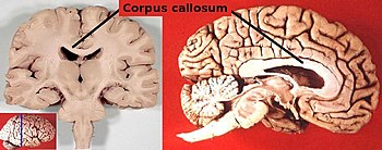
The corpus callosum, a nerve bundle connecting the two cerebral hemispheres, with the lateral ventricles directly below

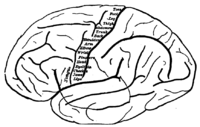
Many of the brain areas Brodmann defined have their own complex internal structures. In a number of cases, brain areas are organized into "topographic maps", where adjoining bits of the cortex correspond to adjoining parts of the body, or of some more abstract entity. A simple example of this type of correspondence is the primary motor cortex, a strip of tissue running along the anterior edge of the central sulcus, shown in the image to the right. Motor areas innervating each part of the body arise from a distinct zone, with neighboring body parts represented by neighboring zones. Electrical stimulation of the cortex at any point causes a muscle-contraction in the represented body part. This "somatotopic" representation is not evenly distributed, however. The head, for example, is represented by a region about three times as large as the zone for the entire back and trunk. The size of a zone correlates to the precision of motor control and sensory discrimination possible[citation needed]. The areas for the lips, fingers, and tongue are particularly large, considering the proportional size of their represented body parts.

In visual areas, the maps are retinotopic—that is, they reflect the topography of the retina, the layer of light-activated neurons lining the back of the eye. In this case too the representation is uneven: the fovea—the area at the center of the visual field—is greatly overrepresented compared to the periphery. The visual circuitry in the human cerebral cortex contains several dozen distinct retinotopic maps, each devoted to analyzing the visual input stream in a particular way[citation needed]. The primary visual cortex (Brodmann area 17), which is the main recipient of direct input from the visual part of the thalamus, contains many neurons that are most easily activated by edges with a particular orientation moving across a particular point in the visual field. Visual areas farther downstream extract features such as color, motion, and shape.

In auditory areas, the primary map is tonotopic. Sounds are parsed according to frequency (i.e., high pitch vs. low pitch) by subcortical auditory areas, and this parsing is reflected by the primary auditory zone of the cortex. As with the visual system, there are a number of tonotopic cortical maps, each devoted to analyzing sound in a particular way.

Within a topographic map there can sometimes be finer levels of spatial structure. In the primary visual cortex, for example, where the main organization is retinotopic and the main responses are to moving edges, cells that respond to different edge-orientations are spatially segregated from one another


Pathology Sources of information

Computed tomography of human brain, from base of the skull to top, taken with intravenous contrast medium.
By placing electrodes on the scalp it is possible to record the summed electrical activity of the cortex, in a technique known as electroencephalography (EEG).[8] EEG measures mass changes in population synaptic activity from the cerebral cortex, but can only detect changes over large areas of the brain, with very little sensitivity for sub-cortical activity. EEG recordings can detect events lasting only a few thousandths of a second. EEG recordings have good temporal resolution, but poor spatial resolution.

Apart from measuring the electric field around the skull it is possible to measure the magnetic field directly in a technique known as magnetoencephalography (MEG).[9] This technique has the same temporal resolution as EEG but much better spatial resolution, although not as good as fMRI. The greatest disadvantage of MEG is that, because the magnetic fields generated by neural activity are very weak, the method is only capable of picking up signals from near the surface of the cortex, and even then, only neurons located in the depths of cortical folds (sulci) have dendrites oriented in a way that gives rise to detectable magnetic fields outside the skull.
A scan of the brain using fMRI
There are several methods for detecting brain activity changes by three-dimensional imaging of local changes in blood flow. The older methods are SPECT and PET, which depend on injection of radioactive tracers into the bloodstream. The newest method, functional magnetic resonance imaging (fMRI), has considerably better spatial resolution and involves no radioactivity.[10] Using the most powerful magnets currently available, fMRI can localize brain activity changes to regions as small as one cubic millimeter. The downside is that the temporal resolution is poor: when brain activity increases, the blood flow response is delayed by 1–5 seconds and lasts for at least 10 seconds. Thus, fMRI is a very useful tool for learning which brain regions are involved in a given behavior, but gives little information about the temporal dynamics of their responses. A major advantage for fMRI is that, because it is non-invasive, it can readily be used on human subjects.

Pathology

A human brain showing frontotemporal lobar degeneration causing frontotemporal dementia

Clinically, death is defined as an absence of brain activity as measured by EEG. Injuries to the brain tend to affect large areas of the organ, sometimes causing major deficits in intelligence, memory, and movement. Head trauma caused, for example, by vehicle or industrial accidents, is a leading cause of death in youth and middle age. In many cases, more damage is caused by resultant edema than by the impact itself. Stroke, caused by the blockage or rupturing of blood vessels in the brain, is another major cause of death from brain damage.

Other problems in the brain can be more accurately classified as diseases than as injuries. Neurodegenerative diseases, such as Alzheimer's disease, Parkinson's disease, motor neurone disease, and Huntington's disease are caused by the gradual death of individual neurons, leading to diminution in movement control, memory, and cognition.

Mental disorders, such as clinical depression, schizophrenia, bipolar disorder and post-traumatic stress disorder may involve particular patterns of neuropsychological functioning related to various aspects of mental and somatic function. These disorders may be treated by psychotherapy, psychiatric medication or social intervention and personal recovery work; the underlying issues and associated prognosis vary significantly between individuals.

Some infectious diseases affecting the brain are caused by viruses and bacteria. Infection of the meninges, the membrane that covers the brain, can lead to meningitis. Bovine spongiform encephalopathy (also known as "mad cow disease") is deadly in cattle and humans and is linked to prions. Kuru is a similar prion-borne degenerative brain disease affecting humans. Both are linked to the ingestion of neural tissue, and may explain the tendency in human and some non-human species to avoid cannibalism. Viral or bacterial causes have been reported in multiple sclerosis and Parkinson's disease, and are established causes of encephalopathy, and encephalomyelitis.

Many brain disorders are congenital, occurring during development. Tay-Sachs disease, Fragile X syndrome, and Down syndrome are all linked to genetic and chromosomal errors. Many other syndromes, such as the intrinsic circadian rhythm disorders, are suspected to be congenital as well. Normal development of the brain can be altered by genetic factors, drug use, nutritional deficiencies, and infectious diseases during pregnancy.

Location of two brain areas that play a critical role in language, Broca's area and Wernicke's area

In human beings, it is the left hemisphere that usually contains the specialized language areas. While this holds true for 97% of right-handed people, about 19% of left-handed people have their language areas in the right hemisphere and as many as 68% of them have some language abilities in both the left and the right hemisphere. The two hemispheres are thought to contribute to the processing and understanding of language: the left hemisphere processes the linguistic meaning of prosody (or, the rhythm, stress, and intonation of connected speech), while the right hemisphere processes the emotions conveyed by prosody.[12] Studies of children have shown that if a child has damage to the left hemisphere, the child may develop language in the right hemisphere instead. The younger the child, the better the recovery. So, although the "natural" tendency is for language to develop on the left, human brains are capable of adapting to difficult circumstances, if the damage occurs early enough.

The first language area within the left hemisphere to be discovered is Broca's area, named after Paul Broca, who discovered the area while studying patients with aphasia, a language disorder. Broca's area doesn't just handle getting language out in a motor sense, though. It seems to be more generally involved in the ability to process grammar itself, at least the more complex aspects of grammar. For example, it handles distinguishing a sentence in passive form from a simpler subject-verb-object sentence — the difference between "The boy was hit by the girl" and "The boy hit the girl."

The second language area to be discovered is called Wernicke's area, after Carl Wernicke, a German neurologist who discovered the area while studying patients who had similar symptoms to Broca's area patients but damage to a different part of their brain. Wernicke's aphasia is the term for the disorder occurring upon damage to a patient's Wernicke's area.

Wernicke's aphasia does not only affect speech comprehension. People with Wernicke's aphasia also have difficulty recalling the names of objects, often responding with words that sound similar, or the names of related things, as if they are having a hard time recalling word associations[citation needed].


Functions of the Esophagus

Illu01 head neck.jpg
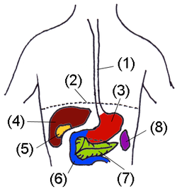
BauchOrgane wn.png

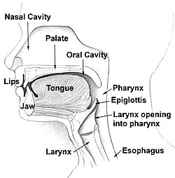
The esophagus or oesophagus (see spelling differences), sometimes known as the gullet, is an organ in vertebrates which consists of a muscular tube through which food passes from the pharynx to the stomach. The word esophagus is derived from the Latin œsophagus, which derives from the Greek word oisophagos (οισοφάγος), lit. "entrance for eating." In humans the esophagus is continuous with the laryngeal part of the pharynx at the level of the C6 vertebra. The esophagus passes through a hole in the level of the tenth thoracic vertebrae (T10). It is usually about 25–30 cm long and connects the mouth to the stomach. It is divided into abdominal parts.